Codominance Examples: Blood Type, Roan & Sickle Cell
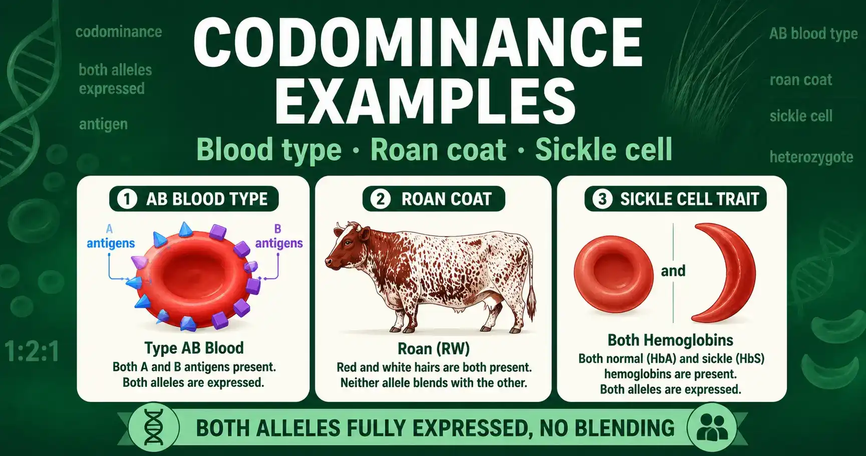
Codominance is an inheritance pattern where both alleles in a heterozygote are fully and separately expressed, with no blending. The three clearest examples are ABO blood type, where a person can show both A and B antigens; roan coat color, where an animal grows both red and white hairs; and sickle cell trait, where the body produces both normal and sickle hemoglobin. In every case, you see both alleles at work at the same time.
This guide walks through each example in depth, with genotype tables and worked Punnett squares you can follow. These cases come up constantly in biology courses and real medicine, from predicting a baby's blood type to understanding why sickle cell carriers are protected against malaria. If you want the broader contrast first, the difference between this pattern and blending is covered in the guide to incomplete dominance vs codominance. Here, the focus is on the examples themselves.
What Makes a Trait Codominant
Codominance means neither allele hides the other, so both appear in the heterozygote at full strength. This sets it apart from complete dominance, where a dominant allele masks a recessive one, and from incomplete dominance, where two alleles blend into an intermediate trait. Under codominance there is no masking and no blending. Both traits simply show up together.
The visual test is straightforward. If you can still pick out each parent's original trait in the offspring, the pattern is codominance. A roan cow has visible red hairs and visible white hairs, not a single pink shade. A person with AB blood has both A markers and B markers on their cells, not a merged "AB-blend" marker. That side-by-side, simultaneous expression is the signature you are looking for in every example below.
One more feature ties these cases together. Because the heterozygote looks different from both homozygous parents, codominant crosses produce a 1:2:1 phenotype ratio rather than the 3:1 of complete dominance. Each genotype maps to its own visible result, which also means you can usually read an organism's genotype straight from its appearance.
Example 1: ABO Blood Type
ABO blood type is the most important codominance example in human genetics, and it adds a second twist: multiple alleles. While any one person carries just two alleles, the population as a whole has three versions of the ABO gene, which sits on chromosome 9. Those three alleles are written IA, IB, and i.
Each allele does something specific. The IA allele puts A antigens on the surface of red blood cells. The IB allele puts B antigens on them. The i allele produces no antigen at all. The IA and IB alleles are codominant with each other, so when both are present, both antigens appear. At the same time, both IA and IB are completely dominant over the recessive i allele. A single gene therefore shows two relationships at once: codominance between IA and IB, and simple dominance over i.
Blood Type Genotypes and Phenotypes
Combining three alleles two at a time gives four possible blood types from six genotypes. The table below shows how each genotype maps to a blood type.
| Blood type (phenotype) | Genotype(s) |
|---|---|
| Type A | IA IA or IA i |
| Type B | IB IB or IB i |
| Type AB | IA IB |
| Type O | ii |
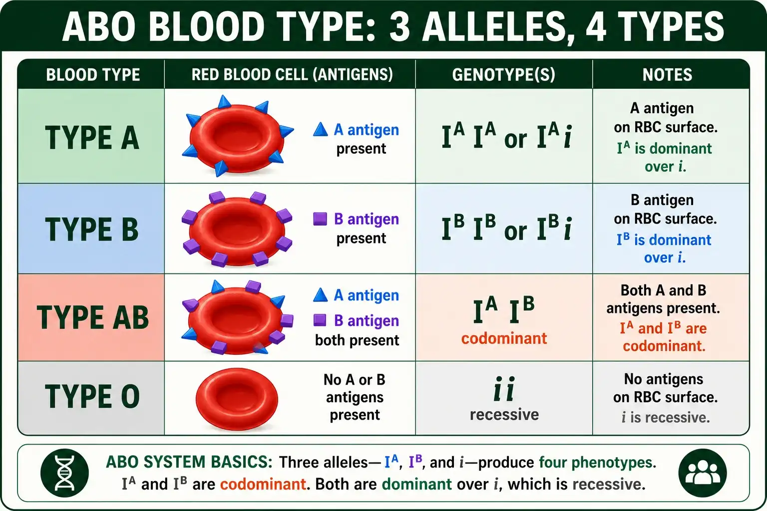
Type AB is the codominance case. An IA IB person makes both A and B antigens, expressing both alleles fully and equally. Type O is the recessive case, requiring two i alleles. Notice that type A and type B each come from two possible genotypes, because the dominant allele shows whether it is paired with a matching allele or with the recessive i. This is exactly why a blood test alone cannot always reveal whether a type A person is IA IA or IA i.
A Worked Blood Type Cross
Consider one of the most striking crosses in genetics: a type A parent and a type B parent who can have children of every blood type. This happens when both parents are heterozygous, with genotypes IA i and IB i.
Work out the gametes. The type A parent (IA i) produces IA and i gametes. The type B parent (IB i) produces IB and i gametes. Fill the grid and the four boxes come out as IA IB, IA i, IB i, and ii. Those translate to type AB, type A, type B, and type O, a 1:1:1:1 ratio across all four blood types.
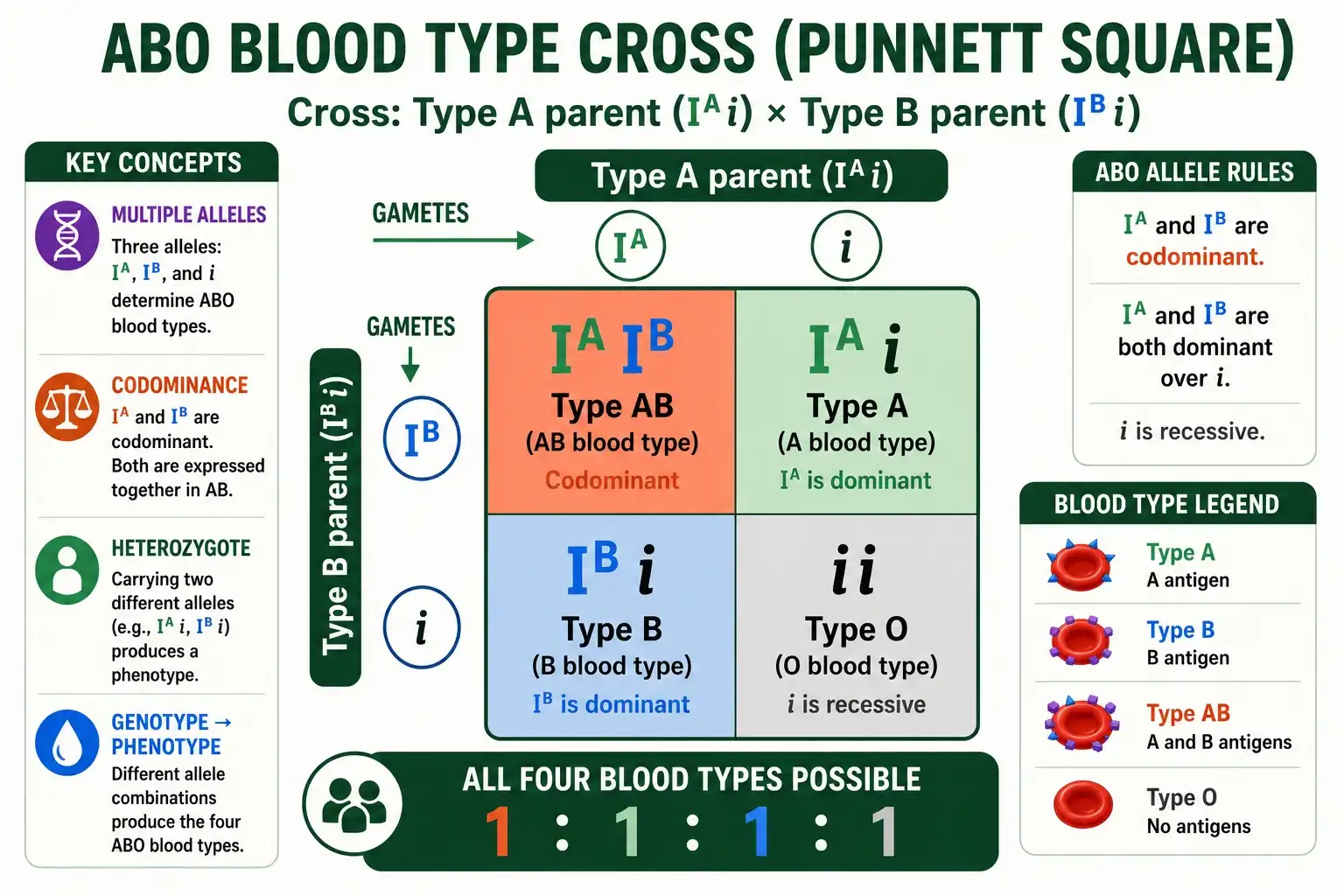
This result surprises people, because two parents with neither AB nor O blood can produce children with both. It also shows how blood typing can rule out parentage. A famous case involved the actor Charlie Chaplin, whose blood type was AB. An AB individual has the genotype IA IB and can only pass on IA or IB, never i. So an AB father cannot have a type O child, since type O requires two i alleles. The science was clear even when a 1940s court ruled otherwise. You can read more about the genetics of these multiple-allele crosses in this Concepts of Biology chapter.
Example 2: Roan Coat Color
Roan coat color in cattle and horses is the cleanest visual example of codominance, because you can literally see both alleles at work. A roan animal carries one allele for red coat and one allele for white coat, and instead of producing a blended pink, the two colors appear together as a mix of red and white hairs.
The key detail is what happens at the level of a single hair. Each hair is either fully red or fully white, never an in-between shade. The roan appearance comes from the two colors being interspersed across the body, so from a distance the animal looks roan, but up close you see distinct red and white hairs. That is co-expression, not blending, which is what makes it codominance rather than incomplete dominance. A blended result would give every hair the same intermediate color.
A roan cross follows the standard codominant pattern. Cross two roan animals, each carrying one red and one white allele, and the offspring come out as one homozygous red, two roan, and one homozygous white, a 1:2:1 ratio. The middle group, the roans, display both coat colors at once.
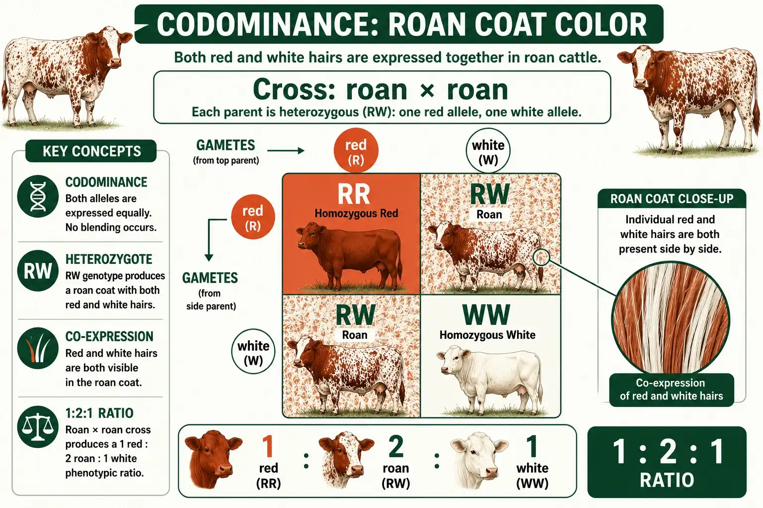
Because each genotype produces a distinguishable coat, breeders can identify an animal's genotype on sight, which is useful in managing herds. A roan animal is always heterozygous, a solid red animal is homozygous red, and a white animal is homozygous white. There is no hidden carrier state, since nothing is masked.
Example 3: Sickle Cell Trait
Sickle cell offers the most scientifically interesting codominance example, because it shows the pattern at the molecular level. The hemoglobin gene has a normal allele, often written HbA, and a sickle allele, written HbS. The sickle allele carries a small mutation that changes the shape of the hemoglobin protein.
The mutation itself is tiny but consequential. A single change in the HBB gene swaps one amino acid in the hemoglobin chain, and that one substitution makes the protein prone to clumping when oxygen is low. The clumping distorts red blood cells into the rigid, curved sickle shape that gives the condition its name. It is a striking reminder that a single-letter change in DNA can ripple all the way up to a serious disease.
There are three genotypes. A person with two normal alleles (HbA HbA) makes only normal hemoglobin and is unaffected. A person with two sickle alleles (HbS HbS) makes only sickle hemoglobin and has sickle cell anemia, a serious disease. The heterozygote (HbA HbS) is the codominant case. Their cells produce both normal hemoglobin and sickle hemoglobin in roughly equal amounts, because both alleles are expressed. This heterozygous state is called sickle cell trait, and it is usually symptom-free under normal conditions.
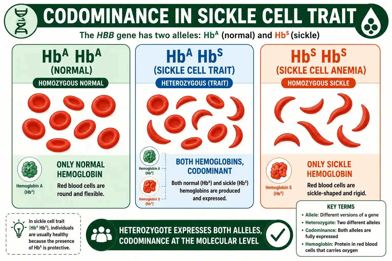
There is a subtlety worth getting right. At the level of the disease, sickle cell behaves like a recessive condition, because you generally need two sickle alleles to develop sickle cell anemia. But at the molecular level, the heterozygote clearly expresses both alleles, producing both protein versions, which is the definition of codominance. So whether sickle cell looks recessive or codominant depends on whether you are looking at the disease or at the proteins in the blood. This is a common point of confusion, and being precise about it is the mark of understanding the example fully.
Sickle cell trait also carries a famous advantage. Carriers, the HbA HbS heterozygotes, are partially protected against malaria. In regions where malaria is widespread, this protection helped the sickle allele persist in the population despite the harm of the homozygous disease. The heterozygous genotype was actually beneficial there, a powerful example of how a single allele copy can be protective. Because each carrier can pass the sickle allele to children, predicting the odds of an affected child is a real concern for families, and a carrier probability calculator is built to estimate exactly that risk.
Example 4: The MN Blood Group
The MN blood group is a tidy, less famous example that shows codominance without the added layer of multiple alleles or dominance over a recessive. It involves just two alleles, often labeled M and N, and they are codominant with each other.
The genotypes are simple. An individual can be MM, MN, or NN. MM individuals carry only the M antigen on their red blood cells, NN individuals carry only the N antigen, and MN individuals carry both antigens at once. The heterozygous MN person expresses both alleles fully, exactly as in the ABO and roan examples. Because there is no recessive allele hiding in the system, every genotype gives a distinct, readable phenotype.
The MN group is useful precisely because it is so clean. With only two codominant alleles and three clearly distinguishable phenotypes, it strips codominance down to its essentials. A cross between two MN parents gives the familiar 1 MM : 2 MN : 1 NN ratio, with the heterozygotes clearly showing both markers. It is a good example to reach for when the ABO system feels complicated by its third allele.
Other Codominance Examples
Several more cases follow the same logic and are worth knowing. Speckled or mottled chickens show codominance when a black-feathered variety and a white-feathered variety produce offspring with both black and white feathers appearing separately, rather than a uniform grey. The distinct patches of color are the giveaway.
Certain flowers display codominance as spotting or striping, where a red-flowered and a white-flowered parent give offspring with both red and white regions on the same petals, instead of pink. Some camellias and azaleas show this pattern. In each of these examples, the rule holds: both parental traits appear at full strength and remain distinguishable, which separates codominance from the blending of incomplete dominance.
Codominance also runs deep in human immunity. The HLA genes, which help the immune system tell your own cells from foreign ones, are codominantly expressed, so you display the markers inherited from both parents at once. This matters enormously for organ and bone marrow transplants, because a donor and recipient need to share enough of these codominant markers for the transplant to succeed. It is the same principle as the blood and coat-color examples, scaled up to one of the most clinically important gene families in the body, which shows how far the reach of a single inheritance pattern can extend.
Codominance and Blood Transfusion Safety
The codominance of the ABO alleles is not just a textbook curiosity. It directly determines which blood transfusions are safe, which makes this one of the most consequential examples of any inheritance pattern.
Your immune system attacks antigens it does not recognize. A person with type A blood carries A antigens and makes antibodies against B. A type B person is the reverse. Because a type AB person expresses both antigens through codominance, they make antibodies against neither, so they can receive blood of any ABO type. This is why type AB is called the universal recipient. Type O, by contrast, carries no A or B antigens at all, so type O blood can be given to almost anyone, making it the universal donor.
Get the match wrong and the consequences are severe. If type B blood is transfused into a type A patient, the recipient's anti-B antibodies attack the donor cells, causing them to clump and break down in a dangerous reaction. Hospitals therefore type blood carefully before any transfusion. In this setting, the codominant expression of the IA and IB alleles is the difference between a safe transfusion and a life-threatening one, which is why the ABO system is studied with such care.
Codominance vs Multiple Alleles: Clearing the Confusion
ABO blood type is often used to illustrate both codominance and multiple alleles, and students sometimes assume the two terms mean the same thing. They do not, and keeping them separate sharpens your understanding of every example.
Multiple alleles describes how many versions of a gene exist in the population. The ABO gene has three, IA, IB, and i, which is more than the two alleles Mendel worked with. This is a statement about variety at the gene level. Codominance, by contrast, describes how two alleles interact when they are paired in one individual. It is a statement about expression in a single heterozygote. The ABO system happens to show both features at once, which is exactly why it is such a popular teaching example.
The distinction becomes clear when you look at other cases. The MN blood group is codominant but has only two alleles, so it shows codominance without multiple alleles. Roan coat color is the same. Meanwhile, a gene could have many alleles in a population that all follow simple dominance, showing multiple alleles without codominance. ABO is special because it combines the two: three alleles in the population, with codominance between IA and IB and simple dominance over i. Recognizing which concept a question is testing keeps your answers precise.
Every codominance example fills a Punnett square the same way as a standard monohybrid cross. The mechanics of combining gametes never change; only the way you label the phenotypes differs, because each genotype keeps its own appearance.
The reliable result is the 1:2:1 phenotype ratio in a cross between two heterozygotes. Cross two roans and you get 1 red : 2 roan : 1 white. Cross two AB individuals and you get 1 type A : 2 type AB : 1 type B, with no type O possible, since neither parent carries the i allele. Cross two MN individuals and you get 1 MM : 2 MN : 1 NN. In each case the two heterozygous boxes show both traits, and the two homozygous boxes show one trait each. If you want to set up and solve any of these crosses step by step, you can start here with the basics of building the grid.
The one habit to keep is reading by genotype first, then translating to phenotype. Because codominant heterozygotes are distinguishable, the genotype ratio and phenotype ratio match at 1:2:1, which makes these crosses cleaner to interpret than complete-dominance crosses where genotypes collapse into shared phenotypes.
Frequently Asked Questions
Is sickle cell codominance or recessive?
Both descriptions apply, depending on the level. At the molecular level, sickle cell trait is codominant, because heterozygotes produce both normal and sickle hemoglobin. At the disease level, sickle cell anemia behaves recessively, since two sickle alleles are usually needed to develop the condition.
Why is AB blood type an example of codominance?
AB blood type is codominance because the IA and IB alleles are both fully expressed. A person with genotype IA IB carries both A antigens and B antigens on their red blood cells at the same time, with no blending into a single intermediate antigen.
Can two type A and type B parents have a type O child?
Yes, if both parents are heterozygous carriers of the recessive i allele. A type A parent (IA i) and a type B parent (IB i) can each pass on i, producing an ii child with type O blood, alongside the chance of type A, B, or AB.
What ratio does a codominant cross produce?
A cross between two heterozygotes produces a 1:2:1 phenotype ratio. Because the heterozygote shows both traits and is visibly distinct from both homozygotes, no genotypes share an appearance, so the phenotype ratio matches the genotype ratio.
Bringing It Together
Codominance shows its hand whenever both alleles appear at full strength in a heterozygote. ABO blood type displays both A and B antigens in type AB individuals and layers in a third allele for type O. Roan coats mix distinct red and white hairs. Sickle cell trait produces both normal and sickle hemoglobin, codominant at the molecular level even though the disease itself is recessive. The MN blood group strips the idea down to two clean codominant alleles.
Across all of them, the same rule applies: no masking, no blending, both traits visible, and a 1:2:1 ratio in a heterozygote cross. A quick way to lock in the pattern is to keep one anchor example in mind for each setting: AB blood for humans, roan coats for animals, and spotted petals for plants. Once those anchors are firm, classifying any new example becomes almost automatic. The fastest way to test any of these crosses and see the genotypes and phenotypes side by side is to lay them out with the Punnett Square Calculator, which handles codominant and multiple-allele patterns alongside the classic Mendelian ones. For a deeper academic treatment of these complex inheritance patterns, this resource is a useful and reliable reference to keep on hand.